Thursday, January 5, 2012

Kelainan Fisiologis Tulang

Mikrosefalus
Mikrosefalus adalah kelainan otak dengan ukuran kepala lebih kecil dari ukuran kepala rata-rata berdasarkan umur dan jenis kelamin. Kepala dikatakan lebih kecil jika ukuran lingkar kepala kurang dari 42 cm atau lebih kecil dari standar deviasi 3 dibawah angka rata-rata.
Mikrosefalus seringkali terjadi akibat kegagalan pertumbuhan otak pada kecepatan yang normal. Beberapa penyakit yang memengaruhi pertumbuhan otak dapat menyebabkan mikrosefalus. Mikrosefalus seringkali berhubungan dengan keterbelakangan mental. Mikrosefalus dapat terjadi setelah infeksi yang menyebabkan kerusakan pada otak pada bayi yang sangat muda (misalnya meningitis dan meningoensefalitis).

·        Penyebab
Mikrosefalus disebabkan oleh gangguan kelainan genetik yang mengganggu pertumbuhan korteks serebral selama bulan-bulan awal perkembangan janin. Hal ini terkait dengan sindrom Down, sindrom kromosom, dan sindrom neurometabolic. Bayi mungkin dilahirkan dengan microcephaly jika selama kehamilan ibu memiliki kebiasaan menyalahgunakan obat-obatan atau alkohol,  terinfeksi cytomegalovirus, virus rubela, teracuni bahan kimia tertentu.

·        Gejala
Gejala – gejala yang muncul pada bayi:
o   Keterbelakangan mental
o   Tertunda fungsi motorik dan bicara
o   Kelainan wajah
o   Perawakan pendek
o   Hiperaktif
o   Kejang
o   Kesulitan dengan koordinasi dan keseimbangan
o   Kelainan neurologis
·        Perawatan
Tidak ada pengobatan untuk microcephaly yang dapat mengembalikan kepala anak ke ukuran normal atau bentuk. Perawatan berfokus pada cara-cara untuk mengurangi dampak neurologis terkait cacat dan cacat. Anak-anak dengan keterlambatan perkembangan microcephaly dan biasanya dievaluasi oleh pediatrik neurolog dan diikuti oleh tim manajemen medis.

Rakitis
Rakitis adalah pelunakan tulang pada anak-anak karena kekurangan atau gangguan metabolisme vitamin D, magnesium, fosfor atau kalsium, berpotensi menyebabkan patah tulang dan kelainan bentuk. Rakitis adalah salah satu penyakit anak yang paling sering di banyak negara berkembang. Penyebab utama adalah kekurangan vitamin D, namun kekurangan kalsium yang memadai dalam diet juga dapat menyebabkan rakitis (kasus diare berat dan muntah dapat menjadi penyebab kekurangan). Meskipun dapat terjadi pada orang dewasa, sebagian besar kasus terjadi pada anak-anak menderita gizi buruk, biasanya akibat kelaparan atau kelaparan selama tahap awal masa kanak-kanak. Osteomalacia adalah istilah yang digunakan untuk menggambarkan kondisi serupa terjadi pada orang dewasa, umumnya karena kekurangan vitamin D. [3] Asal usul "rakitis" kata mungkin dari kata dialek Inggris Kuno 'wrickken', memelintir. Kata berasal Yunani "rachitis" (ραχίτις, yang berarti "peradangan tulang belakang") kemudian diadopsi sebagai istilah ilmiah untuk rakitis, terutama karena kesamaan kata-kata 'dalam suara.

·        Penyebab
Penyebab utama dari rakitis adalah kekurangan vitamin D. Vitamin D diperlukan untuk penyerapan kalsium dari usus. Sinar matahari, sinar ultraviolet terutama, memungkinkan sel-sel kulit manusia mengkonversi vitamin D dari aktif ke keadaan aktif. Dengan tidak adanya vitamin D, kalsium tidak benar diserap, mengakibatkan hipokalsemia, menyebabkan cacat tulang dan gigi dan neuromuskuler gejala, misalnya hyperexcitability. Makanan yang mengandung vitamin D termasuk mentega, telur, minyak hati ikan, margarin, susu dan jus, dan ikan berminyak seperti tuna, ikan herring, dan salmon. Suatu bentuk yang jarang dominan terkait-X ada yang disebut rakitis Vitamin D tahan. Kasus telah dilaporkan di Inggris dalam beberapa tahun terakhir dari rakitis pada anak-anak dari latar belakang sosial yang disebabkan oleh ketidakmampuan untuk membuat vitamin D karena sinar ultraviolet matahari tidak mencapai kulit karena penggunaan terus-menerus dari tabir surya yang kuat, atau terlalu banyak "yang mencakup up "di bawah sinar matahari, atau menghabiskan waktu di dalam ruangan terlalu banyak. Kasus lain telah dilaporkan di antara anak-anak dari beberapa kelompok etnis di mana ibu menghindari paparan matahari untuk alasan agama atau budaya, mengarah ke ibu kekurangan vitamin D. British Medical Journal melaporkan pada 2010 bahwa dokter di Newcastle di Tyne melihat 20 kasus rakitis per tahun.

·        Gejala
Tanda dan gejala rakitis meliputi:
o   Nyeri tulang atau kelembutan
o   Masalah gigi
o   Kelemahan otot (miopati reyot atau "sindrom bayi floppy" atau "bayi Slinky" (di mana bayi floppy atau Slinky-suka)
o   Meningkatkan kecenderungan untuk patah tulang (tulang mudah patah), terutama patah tulang greenstick
o   Deformitas rangka
§  Balita: kaki bengkok (genu varum)
§  Anak lebih tua: Knock-lutut (genu valgum) atau "lutut keanginan"
§  Kranial, tulang belakang, dan panggul cacat
o   Gangguan pertumbuhan
o   Hipokalsemia (tingkat rendah kalsium dalam darah), dan
o   Tetani (kejang otot tidak terkendali di seluruh tubuh).
o   Craniotabes (tengkorak lunak)
o   Costochondral pembengkakan (alias "rosario reyot" atau "rachitic rosario")
o   Harrison alur
o   Malleoli ganda tanda akibat hiperplasia metaphyseal
o   Pelebaran pergelangan tangan menimbulkan kecurigaan awal, itu adalah karena hiperplasia tulang rawan metaphysial.
X-ray atau rontgen dari penderita canggih dari rakitis cenderung hadir dalam cara klasik: kaki busur (kurva luar dari tulang panjang kaki) dan dada cacat. Perubahan dalam tengkorak juga terjadi menyebabkan khas "alun-alun menuju" penampilan. Cacat ini bertahan dalam kehidupan dewasa jika tidak diobati. Konsekuensi jangka panjang termasuk lengkungan permanen atau pengrusakan tulang panjang, dan kembali melengkung.

·        Pengobatan
Rakitis dapat didiagnosis dengan bantuan:
o   Tes darah:
      • Kalsium serum dapat menunjukkan tingkat yang rendah kalsium, fosfor serum mungkin rendah, dan fosfatase alkali serum dapat menjadi tinggi.
o    Gas darah arteri dapat mengungkapkan asidosis metabolik
    • X-ray tulang yang terkena bisa menunjukkan hilangnya kalsium dari tulang atau perubahan bentuk atau struktur tulang.
    • Biopsi tulang jarang dilakukan tetapi akan mengkonfirmasi rakitis.

Pengobatan melibatkan asupan makanan meningkatkan kalsium, fosfat dan vitamin D. Paparan terhadap sinar ultraviolet B (sinar matahari ketika matahari tertinggi di langit), minyak ikan cod, halibut minyak hati, dan viosterol semua sumber vitamin D. Sebuah jumlah yang cukup cahaya ultraviolet B di bawah sinar matahari setiap hari dan persediaan yang memadai kalsium dan fosfor dalam makanan dapat mencegah rakitis. Darker bayi berkulit perlu lebih lama untuk terkena sinar ultraviolet. Penggantian vitamin D telah terbukti benar rakitis menggunakan metode terapi sinar ultraviolet dan obat-obatan. Rekomendasi adalah untuk 400 unit internasional (IU) vitamin D per hari untuk bayi dan anak-anak. Anak-anak yang tidak mendapatkan jumlah yang cukup vitamin D meningkatkan risiko rakitis. Vitamin D sangat penting untuk memungkinkan tubuh untuk penyerapan kalsium untuk digunakan dalam kalsifikasi tulang yang tepat dan pemeliharaan.

Osteoporosis
Osteoporosis adalah penyakit tulang yang mempunyai sifat-sifat khas berupa massa tulang yang rendah, disertai mikro arsitektur tulang dan penurunan kualitas jaringan tulang yang dapat akhirnya menimbulkan kerapuhan tulang.
Klasifikasi:
Osteoporosis primer
Osteoporosis primer sering menyerang wanita paska menopause dan juga pada pria usia lanjut dengan penyebab yang belum diketahui.
Osteoporosis sekunder
Sedangkan osteoporosis sekunder disebabkan oleh penyakit yang berhubungan dengan :
o   Cushing's disease
o   Hyperthyroidism
o   Hyperparathyroidism
o   Hypogonadism
o   Kelainan hepar
o   Kegagalan ginjal kronis
o   Kurang gerak
o   Kebiasaan minum alkohol
o   Pemakai obat-obatan/corticosteroid
o   Kelebihan kafein
o   Merokok
Osteoporosis anak
Osteoporosis pada anak disebut juvenile idiopathic osteoporosis.

·         Penyebab
o   Osteoporosis postmenopausal terjadi karena kekurangan estrogen (hormon utama pada wanita), yang membantu mengatur pengangkutan kalsium ke dalam tulang pada wanita. Biasanya gejala timbul pada wanita yang berusia di antara 51-75 tahun, tetapi bisa mulai muncul lebih cepat ataupun lebih lambat. Tidak semua wanita memiliki risiko yang sama untuk menderita osteoporosis postmenopausal, wanita kulit putih dan daerah timur lebih mudah menderita penyakit ini daripada wanita kulit hitam.
o   Osteoporosis senilis terjadi karena kekurangan kalsium yang berhubungan dengan usia dan ketidakseimbangan di antara kecepatan hancurnya tulang dan pembentukan tulang yang baru. Senilis berarti bahwa keadaan ini hanya terjadi pada usia lanjut. Penyakit ini biasanya terjadi pada usia diatas 70 tahun dan 2 kali lebih sering menyerang wanita. Wanita seringkali menderita osteoporosis senilis dan postmenopausal.
o   Kurang dari 5% penderita osteoporosis juga mengalami osteoporosis sekunder, yang disebabkan oleh keadaan medis lainnya atau oleh obat-obatan.Penyakit ini bisa disebabkan oleh gagal ginjal kronis dan kelainan hormonal (terutama tiroid, paratiroid dan adrenal) dan obat-obatan (misalnya kortikosteroid, barbiturat, anti-kejang dan hormon tiroid yang berlebihan). Pemakaian alkohol yang berlebihan dan merokok bisa memperburuk keadaan ini.
o   Osteoporosis juvenil idiopatik merupakan jenis osteoporosis yang penyebabnya tidak diketahui. Hal ini terjadi pada anak-anak dan dewasa muda yang memiliki kadar dan fungsi hormon yang normal, kadar vitamin yang normal dan tidak memiliki penyebab yang jelas dari rapuhnya tulang.

·        Gejala
Kepadatan tulang berkurang secara perlahan (terutama pada penderita osteoporosis senilis), sehingga pada awalnya osteoporosis tidak menimbulkan gejala. Beberapa penderita tidak memiliki gejala.
Jika kepadatan tulang sangat berkurang sehingga tulang menjadi kolaps atau hancur, maka akan timbul nyeri tulang dan kelainan bentuk.
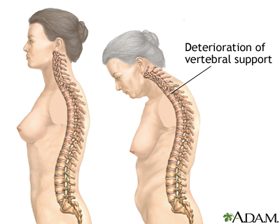
Kolaps tulang belakang menyebabkan nyeri punggung menahun. Tulang belakang yang rapuh bisa mengalami kolaps secara spontan atau karena cedera ringan. Biasanya nyeri timbul secara tiba-tiba dan dirasakan di daerah tertentu dari punggung, yang akan bertambah nyeri jika penderita berdiri atau berjalan. Jika disentuh, daerah tersebut akan terasa sakit, tetapi biasanya rasa sakit ini akan menghilang secara bertahap setelah beberapa minggu atau beberapa bulan. Jika beberapa tulang belakang hancur, maka akan terbentuk kelengkungan yang abnormal dari tulang belakang (punuk Dowager), yang menyebabkan ketegangan otot dan sakit.
Tulang lainnya bisa patah, yang seringkali disebabkan oleh tekanan yang ringan atau karena jatuh. Salah satu patah tulang yang paling serius adalah patah tulang panggul. Yang juga sering terjadi adalah patah tulang lengan (radius) di daerah persambungannya dengan pergelangan tangan, yang disebut fraktur Colles. Selain itu, pada penderita osteoporosis, patah tulang cenderung menyembuh secara perlahan.

·        Pencegahan
Pencegahan osteoporosi meliputi:
o   Mempertahankan atau meningkatkan kepadatan tulang dengan mengonsumsi kalsium yang cukup
o   Melakukan olah raga dengan beban
o   Mengkonsumsi obat (untuk beberapa orang tertentu).
Mengkonsumsi kalsium dalam jumlah yang cukup sangat efektif, terutama sebelum tercapainya kepadatan tulang maksimal (sekitar umur 30 tahun). Minum 2 gelas susu dan tambahan vitamin D setiap hari, bisa meningkatkan kepadatan tulang pada wanita setengah baya yang sebelumnya tidak mendapatkan cukup kalsium. Akan tetapi tablet kalsium dan susu yang dikonsumsi setiap hari akhir - akhir ini menjadi perdebatan sebagai pemicu terjadi osteoporosis, berhubungan dengan teori osteoblast.
Olah raga beban (misalnya berjalan dan menaiki tangga) akan meningkatkan kepadatan tulang. Berenang tidak meningkatkan kepadatan tulang.
Estrogen membantu mempertahankan kepadatan tulang pada wanita dan sering diminum bersamaan dengan progesteron. Terapi sulih estrogen paling efektif dimulai dalam 4-6 tahun setelah menopause; tetapi jika baru dimulai lebih dari 6 tahun setelah menopause, masih bisa memperlambat kerapuhan tulang dan mengurangi risiko patah tulang. Raloksifen merupakan obat menyerupai estrogen yang baru, yang mungkin kurang efektif daripada estrogen dalam mencegah kerapuhan tulang, tetapi tidak memiliki efek terhadap payudara atau rahim. Untuk mencegah osteroporosis, bisfosfonat (contohnya alendronat), bisa digunakan sendiri atau bersamaan dengan terapi sulih hormon.


·        Pengobatan
Pada seseorang yang mengalami patah tulang, diagnosis osteoporosis ditegakkan berdasarkan gejala, pemeriksaan fisik dan rontgen tulang. Pemeriksaan lebih lanjut mungkin diperlukan untuk menyingkirkan keadaan lainnya yang bisa diatasi, yang bisa menyebabkan osteoporosis.
Untuk mendiagnosis osteoporosis sebelum terjadinya patah tulang dilakukan pemeriksaan yang menilai kepadatan tulang. Pemeriksaan yang paling akurat adalah DXA (dual-energy x-ray absorptiometry). Pemeriksaan ini aman dan tidak menimbulkan nyeri, bisa dilakukan dalam waktu 5-15 menit. DXA sangat berguna untuk:
o   Wanita yang memiliki risiko tinggi menderita osteoporosis
o   Penderita yang diagnosisnya belum pasti
o   Penderita yang hasil pengobatannya harus dinilai secara akurat.
Tujuan pengobatan adalah meningkatkan kepadatan tulang. Semua wanita, terutama yang menderita osteoporosis, harus mengonsumsi kalsium dan vitamin D dalam jumlah yang mencukupi.
Wanita paska menopause yang menderita osteoporosis juga bisa mendapatkan estrogen (biasanya bersama dengan progesteron) atau alendronat, yang bisa memperlambat atau menghentikan penyakitnya. Bifosfonat juga digunakan untuk mengobati osteoporosis.
Alendronat berfungsi:
o   Mengurangi kecepatan penyerapan tulang pada wanita pasca menopause
o   Meningkatakan massa tulang di tulang belakang dan tulang panggul
o   Mengurangi angka kejadian patah tulang.
Supaya diserap dengan baik, alendronat harus diminum dengan segelas penuh air pada pagi hari dan dalam waktu 30 menit sesudahnya tidak boleh makan atau minum yang lain. Alendronat bisa mengiritasi lapisan saluran pencernaan bagian atas, sehingga setelah meminumnya tidak boleh berbaring, minimal selama 30 menit sesudahnya. Obat ini tidak boleh diberikan kepada orang yang memiliki kesulitan menelan atau penyakit kerongkongan dan lambung tertentu.
Kalsitonin dianjurkan untuk diberikan kepada orang yang menderita patah tulang belakang yang disertai nyeri. Obat ini bisa diberikan dalam bentuk suntikan atau semprot hidung.
Tambahan fluorida bisa meningkatkan kepadatan tulang. Tetapi tulang bisa mengalami kelainan dan menjadi rapuh, sehingga pemakaiannya tidak dianjurkan.
Pria yang menderita osteoporosis biasanya mendapatkan kalsium dan tambahan vitamin D, terutama jika hasil pemeriksaan menunjukkan bahwa tubuhnya tidak menyerap kalsium dalam jumlah yang mencukupi. Jika kadar testosteronnya rendah, bisa diberikan testosteron.
Patah tulang karena osteoporosis harus diobati. Patah tulang panggul biasanya diatasi dengan tindakan pembedahan. Patah tulang pergelangan biasanya digips atau diperbaiki dengan pembedahan. Pada kolaps tulang belakang disertai nyeri punggung yang hebat, diberikan obat pereda nyeri, dipasang supportive back brace dan dilakukan terapi fisik.

Friday, November 11, 2011

Skin Neoplasm

File:Basal cell carcinoma.jpg
Skin neoplasms (also known as "skin cancer") are skin growths with differing causes and varying degrees of malignancy. The three most common malignant skin cancers are basal cell cancersquamous cell cancer, and melanoma, each of which is named after the type of skin cell from which it arises. Skin cancer generally develops in the epidermis (the outermost layer of skin), so a tumor can usually be seen. This means that it is often possible to detect skin cancers at an early stage. Unlike many other cancers, including those originating in the lungpancreas, and stomach, only a small minority of those affected will actually die of the disease,[1] though it can be disfiguring. Melanoma survival rates are poorer than for non-melanoma skin cancer, although when melanoma is diagnosed at an early stage, treatment is easier and more people survive.[2]  
Skin cancer is the most commonly diagnosed type of cancer. Melanoma and non-melanoma skin cancers combined are more common than lungbreast,colorectal, and prostate cancer.[1] Melanoma is less common than both basal cell carcinoma and squamous cell carcinoma, but it is the most serious — for example, in the UK there were over 11,700 new cases of melanoma in 2008, and over 2,000 deaths.[3] It is the second most common cancer in young adults aged 15–34 in the UK.[4] Most cases are caused by over-exposure to UV rays from the sun or sunbeds.[5] Non-melanoma skin cancers are the most common skin cancers. The majority of these are basal cell carcinomas. These are usually localized growths caused by excessive cumulative exposure to the sun and do not tend to spread.

Classification

There are three main types of skin cancer: basal cell carcinoma (BCC), squamous cell carcinoma (SCC) and malignant melanoma.
CancerDescriptionIllustration
Basal cell carcinomaNote the pearly translucency to fleshy color, tiny blood vessels on the surface, and sometime ulceration which can be characteristics. The key term is translucency.
Basal cell carcinoma3.JPG
Squamous cell carcinomaCommonly presents as a red, crusted, or scaly patch or bump. Often a very rapid growing tumor, pain is a common characteristic.
Squamous Cell Carcinoma1.jpg
Malignant melanomaThe common appearance is an asymmetrical area, with an irregular border, color variation, and often greater than 6 mm diameter.[6]
Melanoma.jpg
Basal cell carcinomas are present on sun-exposed areas of the skin, especially the face. They rarely metastasize and rarely cause death. They are easily treated with surgery or radiation. Squamous cell carcinomas (SCC) are common, but much less common than basal cell cancers. They metastasize more frequently than BCCs. Even then, the metastasis rate is quite low, with the exception of SCCs of the lip, ear, and in immunosuppressed patients. Melanomas are the least frequent of the 3 common skin cancers. They frequently metastasize, and could potentially cause death once they spread.[citation needed]
Less common skin cancers include: Dermatofibrosarcoma protuberansMerkel cell carcinomaKaposi's sarcomakeratoacanthoma, spindle cell tumors, sebaceous carcinomas, microcystic adnexal carcinoma, Pagets's disease of the breast, atypical fibroxanthoma, leimyosarcoma, and angiosarcoma.
The BCC and the SCC often carry a UV-signature mutation indicating that these cancers are caused by UV-B radiation via the direct DNA damage. However the malignant melanoma is predominantly caused by UV-A radiation via the indirect DNA damage.[citation needed] The indirect DNA damage is caused by free radicals and reactive oxygen species. Research indicates that the absorption of three sunscreen ingredients into the skin, combined with a 60-minute exposure to UV, leads to an increase of free radicals in the skin, if applied in too little quantities and too infrequently.[7] However, the researchers add that newer creams often do not contain these specific compounds, and that the combination of other ingredients tends to retain the compounds on the surface of the skin. They also add the frequent re-application reduces the risk of radical formation.

Signs and symptoms

There are a variety of different skin cancer symptoms. These include changes in the skin that do not heal, ulcering in the skin, discolored skin, and changes in existing moles, such as jagged edges to the mole and enlargement of the mole.


Basal cell carcinoma

Basal cell carcinoma usually presents as a raised, smooth, pearly bump on the sun-exposed skin of the headneck or shoulders. Sometimes small blood vessels can be seen within the tumor. Crusting and bleeding in the center of the tumor frequently develops. It is often mistaken for a sore that does not heal. This form of skin cancer is the least deadly and with proper treatment can be completely eliminated, often without scarring.


Squamous cell carcinoma

Squamous cell carcinoma is commonly a red, scaling, thickened patch on sun-exposed skin. Some are firm hard nodules and dome shaped like keratoacanthomas. Ulceration and bleeding may occur. When SCC is not treated, it may develop into a large mass. Squamous cell is the second most common skin cancer. It is dangerous, but not nearly as dangerous as a melanoma.


Melanoma

Most melanomas are brown to black looking lesions. Unfortunately, a few melanomas are pink, red or fleshy in color; these are called amelanotic melanomas. These tend to be more aggressive. Warning signs of malignant melanoma include change in the size, shape, color or elevation of a mole. Other signs are the appearance of a new mole during adulthood or new pain, itching, ulceration or bleeding. An often-used mnemonic is "ABCD", where A= asymmetrical, B= "borders" (irregular= "Coast of Maine sign"), C= "color" (variegated) and D= "diameter" (larger than 6 mm—the size of a pencil eraser).


Other

Merkel cell carcinomas are most often rapidly growing, non-tender red, purple or skin colored bumps that are not painful or itchy. They may be mistaken for a cyst or other type of cancer.[8]


Causes

Ultraviolet radiation from sun exposure is the primary cause of skin cancer.[9][10] Other factors that play a role include:
  • Smoking tobacco[10]
  • HPV infections increase the risk of squamous cell carcinoma.[10]
  • Some genetic syndromes[10] including congenital melanocytic nevi syndrome which is characterized by the presence of nevi (birthmarks or moles) of varying size which are either present at birth, or appear within 6 months of birth. Nevi larger than 20 mm (3/4") in size are at higher risk for becoming cancerous.
  • Chronic non-healing wounds.[10] These are called Marjolin's ulcers based on their appearance, and can develop into squamous cell carcinoma.
  • Ionizing radiation, environmental carcinogens, artificial UV radiation (e.g. tanning beds), aging, and light skin color.[10]


Pathophysiology

Squamous cell carcinoma is a malignant epithelial tumor which originates in epidermis, squamous mucosa or areas of squamous metaplasia.[citation needed]
Macroscopically, the tumor is often elevated, fungating, or may be ulcerated with irregular borders. Microscopically, tumor cells destroy the basement membrane and form sheets or compact masses which invade the subjacent connective tissue (dermis). In well differentiated carcinomas, tumor cells arepleomorphic/atypical, but resembling normal keratinocytes from prickle layer (large, polygonal, with abundant eosinophilic (pink) cytoplasm and central nucleus). Their disposal tends to be similar to that of normal epidermis: immature/basal cells at the periphery, becoming more mature to the centre of the tumor masses. Tumor cells transform into keratinized squamous cells and form round nodules with concentric, laminated layers, called "cell nests" or "epithelial/keratinous pearls". The surrounding stroma is reduced and contains inflammatory infiltrate (lymphocytes). Poorly differentiated squamous carcinomas contain more pleomorphic cells and no keratinization.[11]


Prevention

The risk of developing skin cancer can be reduced through a number of measures including:
  • Decreasing indoor tanning and mid day sun exposure and increasing the use of sunscreen[12]
  • Avoiding the use of tobacco products
  • Reducing overexposure to ultraviolet (UV) radiation, especially in early years
  • Wearing protective clothing (long sleeves and hats) when outdoors
  • Broad-spectrum sunscreen that blocks both UVA and UVB radiation
  • Reapplying sun block according to the manufacturer's directions
There is insufficient evidence to recommend for or against screening for skin cancer,[13]

While sunscreen has been shown to protect against BCC and SCC it may not protect against malignant melanoma. When sunscreen penetrates into the skin it generates reactive chemicals.[7] The experimental and epidemiological evidence suggests that sunscreen use is correlated with malignant melanoma incidence.[14][15][16][17][18][19] This gives rise to questions regarding the possibility that a sunscreen user's lifetime exposure to ultraviolet light may be higher than average. Alternatively, one might question whether sun screens are themselves tumor promoters or carcinogens. Arguably, sunscreen users are the ones most likely to be burned or have been burned by sun light. Similarly, most sunscreens primarily screen UVB, the primary cause of sunburn, while UVA is the primary cause of melanoma. Thus, by limiting the discomfort of sunburn, UVB screening may indirectly result in more UVA exposure. In any case, if some sunscreens promote skin cancer, physical light-scattering sunscreens based in zinc oxide, titanium dioxide or some other natural base are likely safer than chemical blockers such as benzones, etc., as they will be less chemically active.[20]


Management

Treatment is dependent on type of cancer, location of the cancer, age of the patient, and whether the cancer is primary or a recurrence. Treatment is also determined by the specific type of cancer. A small basal cell cancer in a young person the treatment with the best cure rate (Mohs surgery or CCPDMA) might be indicated. In the case of an elderly frail man with multiple complicating medical problems, a difficult to excise basal cell cancer of the nose might warrant radiation therapy (slightly lower cure rate) or no treatment at all. Topical chemotherapy might be indicated for large superficial basal cell carcinoma for good cosmetic outcome, whereas it might be inadequate for invasive nodular basal cell carcinoma or invasive squamous cell carcinoma.[citation needed]. In general, melanoma is poorly responsive to radiation or chemotherapy.
For low-risk disease, radiation therapy (external beam radiotherapy or brachytherapy), topical chemotherapy (imiquimod or 5-fluorouracil) and cryotherapy (freezing the cancer off) can provide adequate control of the disease; both, however, may have lower overall cure rates than certain type of surgery. Other modalities of treatment such as photodynamic therapy, topical chemotherapy,electrodesiccation and curettage can be found in the discussions of basal cell carcinoma and squamous cell carcinoma.
Mohs' micrographic surgery (Mohs surgery) is a technique used to remove the cancer with the least amount of surrounding tissue and the edges are checked immediately to see if tumor is found. This provides the opportunity to remove the least amount of tissue and provide the best cosmetically favorable results. This is especially important for areas where excess skin is limited, such as the face. Cure rates are equivalent to wide excision. Special training is required to perform this technique. An alternative method is CCPDMA and can be performed by a pathologist not familiar with Mohs surgery.
In the case of disease that has spread (metastasized), further surgical procedures or chemotherapy may be required.[21]


Reconstruction

Currently, surgical excision is the most common form of treatment for skin cancers. The goal of reconstructive surgery is restoration of normal appearance and function. The choice of technique in reconstruction is dictated by the size and location of the defect. Excision and reconstruction of facial skin cancers is generally more challenging due to presence of highly visible and functional anatomic structures in the face.
When skin defects are small in size, most can be repaired with simple repair where skin edges are approximated and closed with sutures. This will result in a linear scar. If the repair is made along a natural skin fold or wrinkle line, the scar will be hardly visible. Larger defects may require repair with a skin graft, local skin flap, pedicled skin flap, or a microvascular free flap. Skin grafts and local skin flaps are by far more common than the other listed choices.
Skin grafting is patching of a defect with skin that is removed from another site in the body. The skin graft is sutured to the edges of the defect, and a bolster is placed atop the graft for seven to ten days, to immobilize the graft as it heals in place. There are two forms of skin grafting: split thickness and full thickness. In a split thickness skin graft, a shaver is used to shave a layer of skin from the abdomen or thigh. The donor site, regenerates skin and heals over a period of two weeks. In a full thickness skin graft, a segment of skin is totally removed and the donor site needs to be sutured closed.[22] Split thickness grafts can be used to repair larger defects, but the grafts are inferior in their cosmetic appearance. Full thickness skin grafts are more acceptable cosmetically. However, full thickness grafts can only be used for small or moderate sized defects.
Local skin flaps are a method of closing defects with tissue that closely matches the defect in color and quality. Skin from the periphery of the defect site is mobilized and repositioned to fill the deficit. Various forms of local flaps can be designed to minimize disruption to surrounding tissues and maximize cosmetic outcome of the reconstruction. Pedicled skin flaps are a method of transferring skin with an intact blood supply from a nearby region of the body. An example of such reconstruction is a pedicled forehead flap for repair of a large nasal skin defect. Once the flap develops a source of blood supply form its new bed, the vascular pedicle can be detached.[23]


Prognosis

The mortality rate of basal cell and squamous cell carcinoma are around 0.3% causing 2000 deaths per year in the US. In comparison the mortality rate of melanoma is 15-20% and it causes 6500 deaths per year.[24]:29,31 Even though it is much less common, malignant melanoma is responsible for 75% of all skin cancer-related deaths.[25]


Epidemiology


Age-standardized death from melanomaand other skin cancers per 100,000 inhabitants in 2004.[26]
  no data
  less than 0.7
  0.7-1.4
  1.4-2.1
  2.1-2.8
  2.8-3.5
  3.5-4.2
  4.2-4.9
  4.9-5.6
  5.6-6.3
  6.3-7
  7-7.7
  more than 7.7
A study of the incidence of non-melanoma skin cancer from 1992 to 2006 in the United States was performed by the dermatologist Howard Rogers, MD, PhD, and his colleagues based on the evaluation of Medicare databases. The results of their research showed that cases of non-melanoma skin cancer rose an average of 4.2% a year.[27]
More than 3.5 million cases of skin cancer are diagnosed annually in the United States, which makes it the most common form of cancer in that country. According to the Skin Cancer Foundation, one in five Americans will develop skin cancer at some point of their lives. The first most common form of skin cancer is basal cell carcinoma, followed by the squamous cell carcinoma. Although the incidence of many cancers in the United States is falling, the incidence of melanoma keeps growing, with approximately 68,729 melanomas diagnosed in 2004 according to reports of the National Cancer Institute.[28]
The survival rate for patients with melanoma depends upon when they start treatment. The cure rate is very high when melanoma is detected in early stages, when it can easily be removed surgically. The prognosis is less favorable if the melanoma has spread to other parts of the body.[29]
In the UK, 84,500 non-melanoma skin cancers were registered in 2007 although a study estimated that at least 100,000 cases are diagnosed each year. Most NMSCs were basal cell carcinomas or squamous cell carcinomas. In 2007, 10,672 cases of malignant melanoma were diagnosed.[30]
According to the British Association of Dermatologists children, from 0 to 14 years, and teenagers, from 15 to 19 years, exhibit the highest rates of skin cancers of any European country. Furthermore, incidence of melanoma increased four times in UK teenagers from 1978 to 1997.[31]
Australia exhibits one of the highest rates of skin cancer incidence in the world, almost four times the rates registered in the United States, the UK andCanada. Around 434,000 people receive treatment for non-melanoma skin cancers and 10,300 are treated for melanoma. Melanoma is the common type of cancer in people between 15–44 years in Australia.[32]